A case from Dr Chandramouli’s electrophysiology lab.
A 27-year-old man walks in with a heart rate of 49. In most young adults, the fix is routine: one pacing lead in the ventricle, done in under an hour. In this man, the routine fix could slowly damage the very chamber keeping him alive.
He was born with congenitally corrected transposition of the great arteries (CCTGA). His ventricles are swapped. The chamber built for low-pressure work has pumped blood to his whole body since birth, and the wiring that runs through it follows a route found in almost no other heart.
This is the story of how the team found that hidden wiring with 3D mapping, placed a single lead on it, and kept his heartbeat as narrow after pacing as it was before: 120 ms at baseline, 126 ms on the day-one paced ECG.
Case at a glance
- Patient: 27-year-old man with congenitally corrected transposition of the great arteries (CCTGA)
- Problem: Atrial arrhythmia with slow ventricular response, 49/min, narrow QRS of 120 ms
- Treatment: 3D-mapped conduction system pacing with a single lead and a single-chamber VVI pacemaker
- Result on day one: Paced at 80/min, QRS 126 ms, capture threshold 0.62 V at 0.4 ms
Click any image to open it full size.
The patient: a slow pulse in a rearranged heart
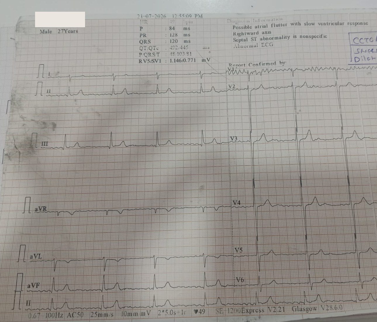
The patient is a 27-year-old man with known CCTGA. His 12-lead ECG on 21 July 2026 showed a ventricular rate of 49 beats per minute. The atria were not in normal sinus rhythm: the machine read “possible atrial flutter with slow ventricular response”, and the tracing was clinically read as an atrial arrhythmia with slow ventricular response.
Two details on this ECG shaped everything that followed.
- The rate is slow. A fast atrial rhythm should drive the ventricles quickly. A ventricular rate of 49 means the AV conduction system is letting very few impulses through. The junction box is failing.
- The QRS is narrow, at 120 ms. Whatever impulses do get through still travel down the heart’s own fast wiring. Both ventricles contract together. That synchrony is an asset worth protecting.
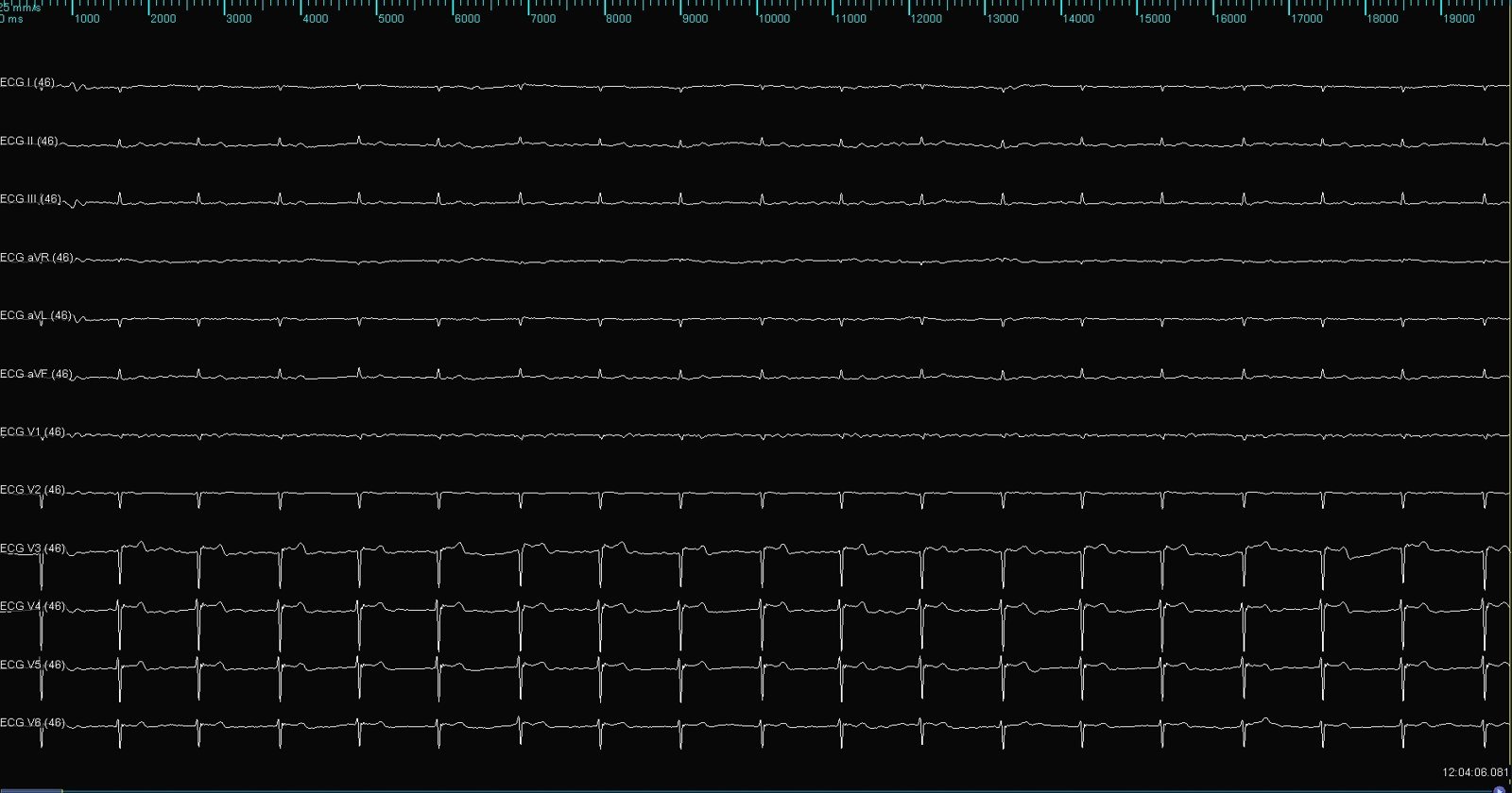
On the table, before any catheter was moved, the electrophysiology recording system confirmed the same picture: slow, narrow-complex ventricular beats.
| Baseline finding | Value |
|---|---|
| Age, sex | 27 years, male |
| Diagnosis | CCTGA |
| Atrial rhythm | Atrial arrhythmia (machine read: possible atrial flutter) |
| Ventricular rate | 49/min |
| QRS duration | 120 ms |
| QT / QTc | 422 / 445 ms |
Understanding CCTGA: two wrongs that make a fragile right
In CCTGA the heart is connected wrongly twice, and the two errors cancel out. The right atrium empties into a morphological left ventricle, which pumps to the lungs. The left atrium empties into a morphological right ventricle, which pumps to the body. Blood goes to the right places, so many patients feel well for decades.
The price is paid in two ways.
The systemic pump is the wrong ventricle. A right ventricle is built for the low-pressure lung circuit. In CCTGA it does high-pressure work for life. It is prone to dilating, weakening and leaking through its tricuspid valve over time.
The wiring takes a detour. Because the atrial and ventricular septa are misaligned, the usual posterior AV node is underdeveloped and cannot connect to the bundle. A second, anterior AV node takes over. It sits below the opening of the right atrial appendage, and its His bundle runs a long course under the pulmonary valve before descending along the septum (Baruteau et al., J Am Heart Assoc 2017).
A long, stretched bundle is a fragile bundle. The same review reports that about 10% of people with CCTGA first present with complete AV block, that the risk of new AV block is roughly 2% per year, and that 30% to 38% of older children and adults have complete AV block. A slow pulse in a 27-year-old with CCTGA is the expected natural history arriving on schedule.
The pacing dilemma: why the routine lead was the wrong lead
A transvenous pacing lead in CCTGA does not land in a right ventricle. It follows the veins into the subpulmonary chamber, which here is the morphological left ventricle. Pacing from its apex makes the two ventricles contract out of step.
In a normal heart that dyssynchrony is tolerated for years. In CCTGA it lands on a systemic right ventricle with little reserve. The Baruteau review links single-site subpulmonary pacing to deterioration of the systemic right ventricle and worsening of the systemic AV valve leak. It cites one series of 53 paced patients in which 14 (26%) later needed an upgrade to biventricular pacing for systemic ventricular dysfunction.
This patient would be paced close to 100% of the time, for an expected five or more decades. Three options were on the table.
| Option | What it does | Problem in this patient |
|---|---|---|
| Conventional apical lead | Paces muscle directly; wide QRS | Dyssynchrony on a systemic right ventricle, for life |
| Biventricular pacing (CRT) | Two ventricular leads resynchronise the walls | Coronary venous anatomy is unpredictable in CCTGA; more hardware in a 27-year-old |
| Conduction system pacing (CSP) | One lead on the His-Purkinje system; near-normal activation | The bundle sits in an unusual place and must be found first |
His own conduction below the block was still healthy, as the narrow 120 ms QRS showed. CSP could hand the impulse straight back to that wiring. The team chose CSP, and solved its one problem, finding the bundle, with 3D mapping.
The procedure: map first, then pace
The procedure took place on 1 September 2026. It ran in four steps.
Step 1: Find the His bundle
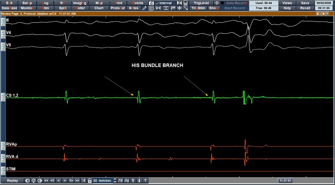
In a normal heart the operator knows where the His bundle is before the first X-ray. In CCTGA the landmarks do not apply. The team advanced a multi-electrode high-density mapping catheter into the subpulmonary ventricle and hunted for the sharp, brief His potential.
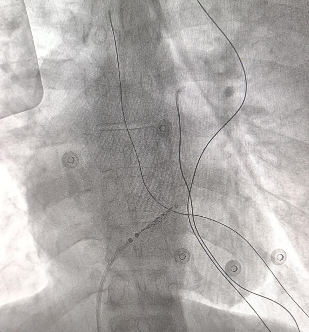
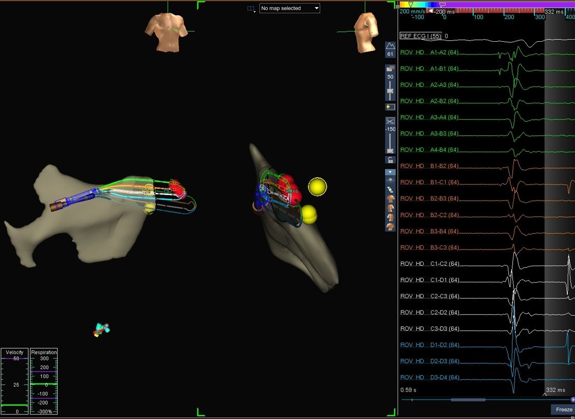
Each point where a His potential appeared was tagged on a 3D electroanatomical map. The result was a cluster of markers showing exactly where this patient’s conduction system runs.
Step 2: Steer the pacing lead to the target
With the target marked, the pacing lead (Abbott UltiPace) was itself displayed on the 3D map as the green catheter. The operator could watch the lead tip travel toward the tagged site in three dimensions, and confirm on the lead’s own signal that it had arrived. The site is marked “CSP” on the map.
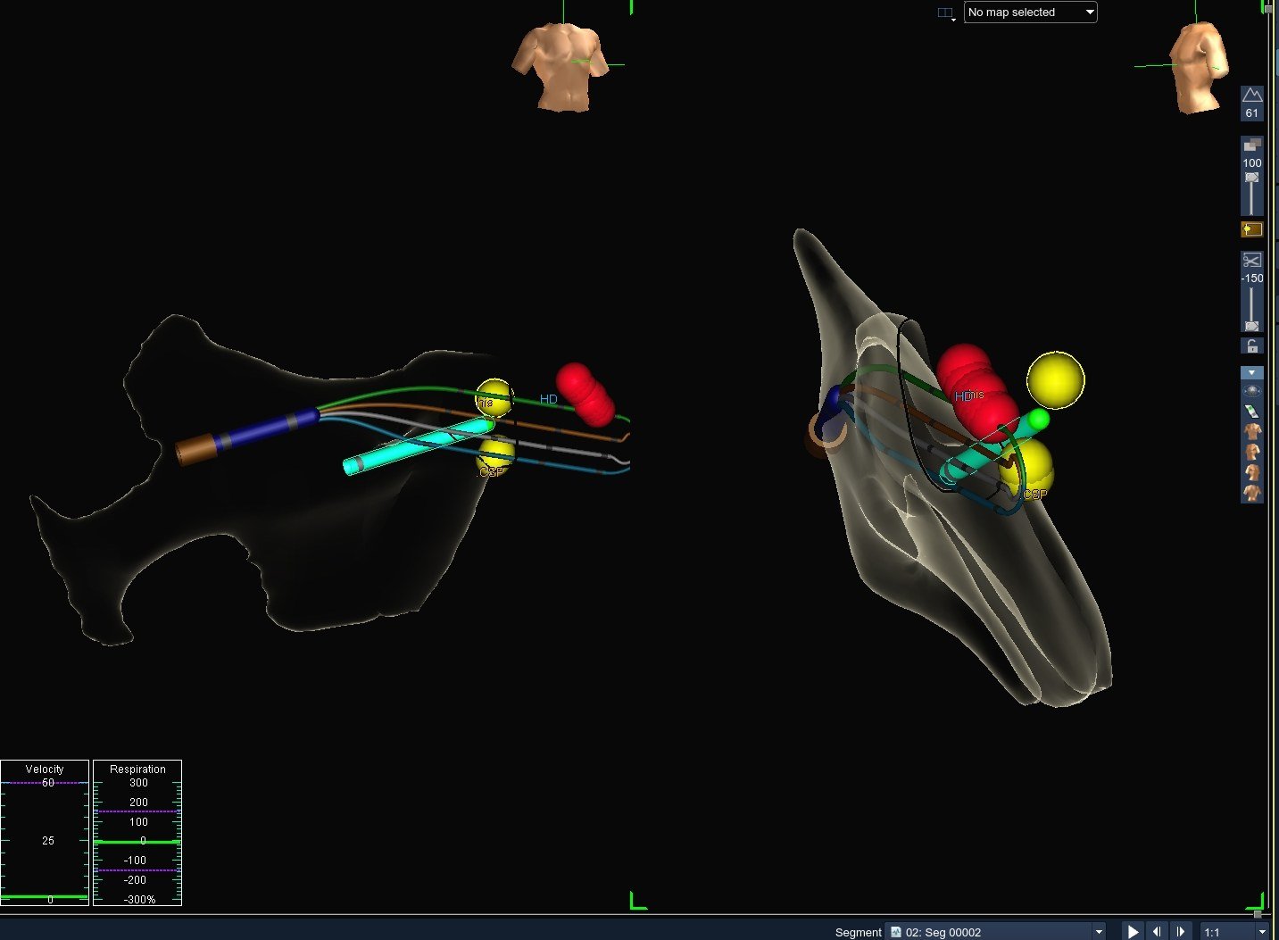
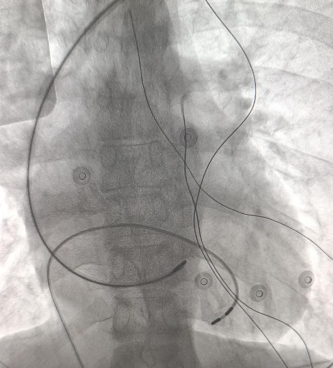
Mapping with the lead visible in 3D turns a blind search into a guided delivery. That matters most in a heart where no textbook picture applies.
Step 3: Radiofrequency ablation, then check the paced beat
Radiofrequency ablation was performed during the same sitting. The ECG afterwards shows every beat paced, at a regular rate, with a QRS of 123 ms.
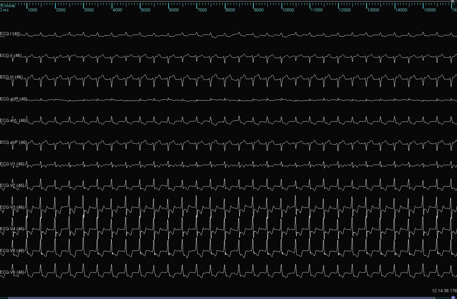
The baseline QRS was 120 ms. A paced QRS of 123 ms means the lead is recruiting the conduction system, not just nearby muscle.
Step 4: Final position and implant numbers
A single-chamber MRI-conditional pacemaker (Abbott Zenus MRI) was connected and placed in the left chest. With a permanent atrial arrhythmia there is no organised atrial beat to track, so a single ventricular lead in VVI mode is the logical system.
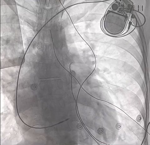
The outcome: one day later
On 2 September 2026, the morning after implant, the 12-lead ECG showed a ventricular paced rhythm at 80 beats per minute with a QRS of 126 ms. The heart rate had gone from 49 to 80. The QRS had moved by 6 ms.
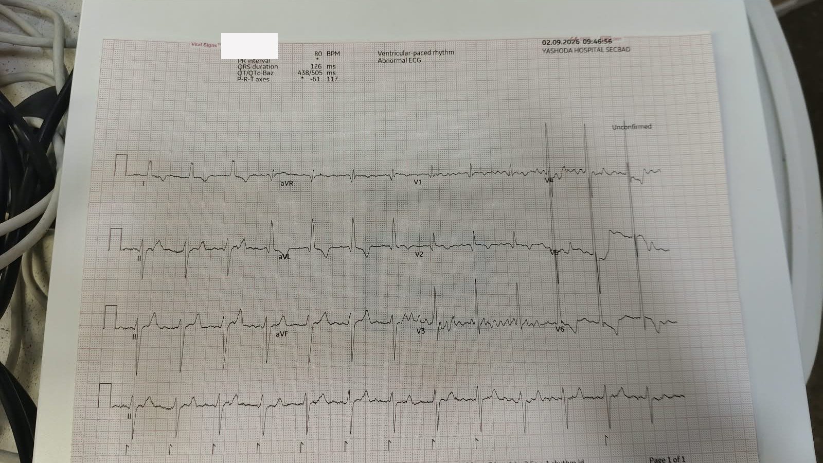
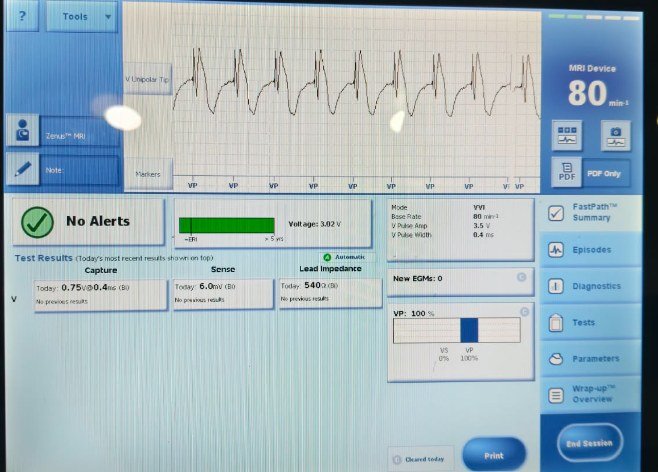
The device check told the same story. Every number was stable or better than at implant, and the pacemaker reported no alerts.
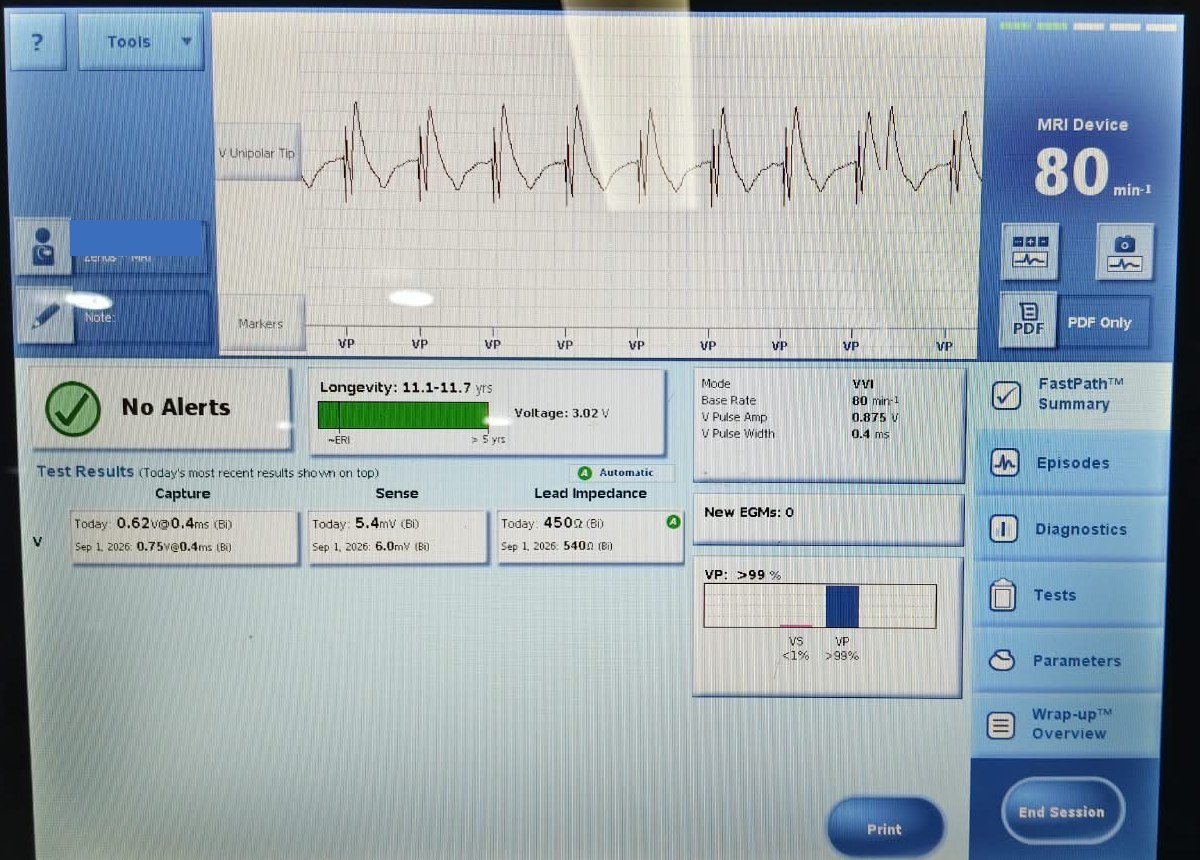
| Parameter | Implant, 1 Sep 2026 | Day one, 2 Sep 2026 |
|---|---|---|
| Mode and base rate | VVI, 80/min | VVI, 80/min |
| Capture threshold (bipolar) | 0.75 V at 0.4 ms | 0.62 V at 0.4 ms |
| Sensed R wave | 6.0 mV | 5.4 mV |
| Lead impedance | 540 ohms | 450 ohms |
| Programmed output | 3.5 V at 0.4 ms | 0.875 V at 0.4 ms |
| Ventricular pacing | 100% | Over 99% |
| Estimated battery longevity | Not shown | 11.1 to 11.7 years |
The threshold deserves a second look. Conduction system leads have a reputation for needing more energy than ordinary leads. Here the lead captured at 0.62 V, which let the output be programmed below 1 V. For a patient who will depend on pacing for life, low output means fewer generator changes.
| ECG | Rhythm | Rate | QRS |
|---|---|---|---|
| Baseline, 21 Jul 2026 | Atrial arrhythmia, slow ventricular response | 49/min | 120 ms |
| After ablation, on table | Paced | Regular paced rate | 123 ms |
| Day one, 2 Sep 2026 | Ventricular paced | 80/min | 126 ms |
Discussion: what this case teaches
The paced QRS is the result that matters. A conventional lead in this ventricle would typically produce a QRS well above 150 ms. In the largest published series of CSP in CCTGA, a 10-centre collaborative study of 15 patients, the paced QRS was 112 ms with conduction system pacing against 164 ms with conventional ventricular pacing (Moore et al., Heart Rhythm 2020). This patient’s 126 ms sits with the first group.
The implant numbers match published experience. That series reported a median threshold of 0.5 V and a median R wave of 6 mV at implant, with acute success in 13 of 15 patients and no lead dysfunction over a median of 8 months. This case: 0.75 V falling to 0.62 V, and an R wave of 6.0 mV.
3D mapping changes the difficulty of the procedure. The hard part of CSP in CCTGA is not fixing the lead. It is knowing where to aim. Tagging His potentials first and then showing the lead on the same map removes the guesswork, and it should cut fluoroscopy time in a young patient.
The anatomy that causes the block may also help the fix. The authors of the collaborative study suggest that the unusual anatomy of CCTGA may favour CSP over conventional cardiac resynchronisation. The bundle is long and reachable from the subpulmonary ventricle, and it delivers the impulse to both ventricles at once.
What this case cannot yet tell us. One day of follow-up proves the lead works. It does not prove the systemic right ventricle is protected. That answer comes from serial echocardiograms, thresholds at 3 and 12 months, and years of follow-up. The published evidence is also small: 15 patients, retrospective, with a median follow-up of 8 months.
Key takeaways
For clinicians
- A slow ventricular rate in a young adult with CCTGA is conduction disease until proven otherwise. The risk of new AV block is about 2% per year.
- Before choosing a lead position, ask which ventricle is systemic and how long the patient will be paced.
- A narrow baseline QRS means the distal conduction system is intact. That makes CSP a strong option.
- Map the His bundle in 3D before delivering the lead. Do not rely on normal-heart landmarks.
- Judge success on the paced QRS and the threshold together. Here: 126 ms and 0.62 V.
For patients and families
- CCTGA can stay silent for years. A slow pulse, tiredness, breathlessness or fainting needs a heart rhythm review.
- Not all pacemakers work the same way. Where the lead sits decides how naturally the heart contracts.
- Conduction system pacing uses the heart’s own wiring, so both pumping chambers beat together.
- Lifelong follow-up with a team that knows congenital heart disease matters as much as the procedure.
References
- Baruteau AE, et al. Cardiac Conduction System in Congenitally Corrected Transposition of the Great Arteries and Its Clinical Relevance. J Am Heart Assoc. 2017.
- Moore JP, et al. Permanent conduction system pacing for congenitally corrected transposition of the great arteries: a PACES/ISACHD collaborative study. Heart Rhythm. 2020.
This article is for education. It is not medical advice for any individual patient.