When an Irregular Heart Rhythm Needs More Than Medicines
by –
Dr. M. S. Chandramouli, MD, DM, IDCC
Consultant Cardiac Electrophysiologist, Department of Cardiology
Yashoda Hospitals, Secunderabad
Atrial fibrillation, often called AFib or AF, can be frightening because it changes something people usually take for granted: the steady rhythm of the heart. A normal heartbeat is organised and predictable. In AFib, the upper chambers of the heart do not squeeze in a coordinated way. Instead, they quiver with fast, disorganised electrical signals. The result can be an irregularly irregular pulse, palpitations, breathlessness, tiredness, dizziness, chest discomfort, reduced exercise capacity, or sometimes no symptoms at all.
For many patients, AFib is not just a “fast heartbeat”. It can affect daily confidence. Some people stop walking longer distances because they fear breathlessness. Others feel exhausted despite normal routines. Some are told about AFib only after an ECG, Holter monitor, smartwatch alert, or routine health check. Because the rhythm may come and go in the early stages, the diagnosis often requires both patient history and rhythm documentation.
The important message is this: AFib is common, especially with age and conditions such as high blood pressure, diabetes, obesity, thyroid disease, sleep apnoea, alcohol excess and other heart diseases. But it is also treatable. The right care plan usually has three goals – control the rhythm or heart rate, reduce stroke risk where needed, and treat the underlying factors that make the rhythm more likely to return.
AFib treatment is not only about slowing the pulse. It is about protecting quality of life, reducing risk, and restoring rhythm confidence wherever possible.
Understanding AFib in Simple Words
The heart runs on electricity. With each beat, an electrical signal travels through the heart in a sequence that allows the chambers to contract efficiently. In atrial fibrillation, multiple chaotic signals arise in the upper chambers, especially near the pulmonary veins – the veins that bring oxygen-rich blood from the lungs into the left atrium. When these triggers repeatedly disturb the heart, the atria may quiver instead of squeezing properly.
This explains why patients may feel a racing heartbeat, an uneven pulse, breathlessness or fatigue. It also explains why AFib is often managed by a heart-rhythm specialist, also called a cardiac electrophysiologist. The specialist evaluates the rhythm pattern, symptom burden, stroke risk, heart structure and triggers before deciding whether medicines, blood thinners, lifestyle correction, cardioversion, catheter ablation or a combination is best.
Modern electrophysiology has changed how AFib is treated. Instead of relying only on medicines, many patients can now be considered for catheter-based treatment. In catheter ablation, thin flexible tubes are passed through a vein in the leg to reach the heart. There are no surgical cuts on the chest. The goal is to electrically isolate the areas that trigger AFib and, when needed, treat additional rhythm circuits that are sustaining the arrhythmia.
Where Pulsed Field Ablation Fits In
Pulsed Field Ablation, or PFA, is one of the most important recent advances in AFib ablation. Traditional ablation methods use heat, as in radiofrequency ablation, or cold, as in cryoballoon ablation. PFA is different. It uses very short, high-energy electrical pulses to affect heart-muscle cells through a process called irreversible electroporation. In simple terms, it targets the electrical trigger tissue without depending on heat or freezing.
One of the reasons PFA has generated interest is tissue selectivity. In carefully performed procedures, PFA is designed to preferentially act on heart muscle while being relatively gentle on nearby structures. This is especially relevant because the left atrium sits close to important surrounding tissues. PFA can also be efficient, which can help streamline pulmonary vein isolation in suitable patients.
However, PFA is not a one-size-fits-all answer. Some patients have AFib alone; others have AFib along with atrial flutter or a more complex scar-related circuit. In those situations, the electrophysiologist may combine technologies. PFA may be used to isolate pulmonary vein triggers, while radiofrequency ablation may be used point-by-point to treat a mapped flutter circuit. The case below is an example of that combined strategy.
Figure 1. High-density 3-D activation mapping. The coloured timing map and intracardiac electrograms help the electrophysiology team understand how the electrical wave is moving through the left atrium.
The Case: Persistent AFib with an Additional Left Atrial Flutter
This clinical case involved an 84-year-old gentleman who had symptomatic persistent atrial fibrillation for about three months. During the procedure, the team also identified a second rhythm problem: left atrial flutter. This matters because AFib and flutter are related but not identical. AFib is usually chaotic and irregular, while atrial flutter is often a more organised re-entrant circuit, where the electrical wave travels repeatedly around a pathway.
For a patient, both can feel like palpitations, fatigue, breathlessness or exercise limitation. For the electrophysiology team, they require careful mapping because the treatment target may be different. If only the pulmonary veins are isolated but an active flutter circuit remains, the patient may continue to have rhythm symptoms. That is why this case was planned as a single-sitting combined approach: pulsed field ablation for pulmonary vein isolation and radiofrequency ablation for the left atrial flutter.
The key message is not that every elderly patient needs the same procedure. The message is that age alone should not be the only deciding factor. A well-selected older patient, assessed by an experienced team, may still benefit from advanced rhythm treatment when symptoms and rhythm findings support it.
Seeing the Rhythm in 3-D
A major strength of modern electrophysiology is that doctors do not have to depend only on X-ray shadows. They can build a three-dimensional electroanatomical model of the patient’s heart chamber in real time. Catheters with multiple electrodes collect thousands of points. The system then displays colour-coded maps that show the timing and voltage of electrical signals.
In this case, high-density mapping was used with a multielectrode HD Grid catheter, a coronary-sinus catheter and the PFA catheter. The activation map showed how the electrical wave moved through the left atrium. The voltage map helped identify relatively healthy tissue and lower-voltage areas that could be related to abnormal conduction. Together, these maps guided the strategy: isolate the pulmonary veins and then identify the critical isthmus of the left atrial flutter circuit.
VIDEO 1. Place the first uploaded MP4 here on the website: dynamic 3-D activation mapping / LAT isochronal movement. In DOCX/PDF this is shown as a static thumbnail.
Step 1: Pulmonary Vein Isolation with PFA
Most AFib begins from triggers around the pulmonary veins. Pulmonary vein isolation, often abbreviated as PVI, is therefore the foundation of many AFib ablation procedures. The aim is to electrically separate the pulmonary vein trigger areas from the rest of the left atrium so that abnormal signals cannot repeatedly start AFib.
In this case, wide antral pulmonary vein isolation was performed using pulsed field energy. “Wide antral” means the treatment is delivered around the vein openings at a safe and strategic distance rather than only at the narrow mouth of the veins. The 3-D map helped the team understand where the catheter was, where energy was delivered, and how the electrical effect changed the rhythm environment.
For a patient reading this, the key reassurance is that catheter ablation is a keyhole procedure. The catheters are typically introduced through the groin vein and guided to the heart. The chest is not opened. Sedation or anaesthesia is used for comfort. The exact duration and hospital stay vary depending on the rhythm complexity and patient condition.
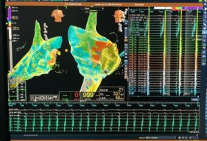
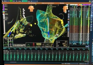
Figure 2. Activation map with lesion tags. The yellow/white tag markers show areas treated around pulmonary-vein antra and mapped regions in the left atrium.
Step 2: Mapping the Left Atrial Flutter Circuit
After addressing the AFib trigger zone, the team characterised the concomitant left atrial flutter. This part of the case is important because atrial flutter often depends on a critical pathway or isthmus. If the isthmus is accurately identified, radiofrequency ablation can be used point-by-point to interrupt the circuit.
The map used local activation timing, also called LAT, to show the order in which areas activated. Where the colour bands crowd together, it can suggest slow conduction – a clue to the pathway sustaining the flutter. The electrophysiologist then targets the critical isthmus rather than treating randomly. This is the value of 3-D mapping: it makes complex rhythm behaviour visible and actionable.
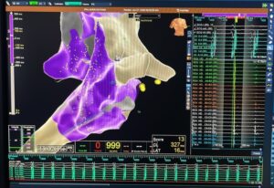
Figure 3. Re-entrant circuit definition. Crowding of isochrones on the LAT map helped localise the slow-conducting isthmus that was subsequently targeted.
Step 3: Radiofrequency Ablation for the Flutter
PFA was used for the AFib component, but the left atrial flutter required a different precision tool. Radiofrequency ablation uses controlled heat to create small, targeted lesions. In this case, point-by-point RFA was applied across the critical isthmus until the flutter was interrupted.
This combination is a practical lesson for patients: advanced care is not about one machine alone. It is about choosing the right energy source for the right target. PFA can be excellent for pulmonary vein isolation in appropriate AFib cases, while RFA remains valuable for mapped flutter circuits and other point-specific targets.
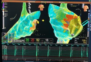
Figure 4. Radiofrequency ablation during the left atrial flutter component. The mapping screen displays power, temperature, impedance, RF time, cycle length and local activation timing during active treatment.
The Final Check: Confirming the Result
A procedure is not complete simply because energy has been delivered. The team must confirm the result. In this case, a completed activation model was re-acquired after treatment. This helped confirm that the targeted rhythm mechanism had been addressed before catheters were withdrawn.
The case documentation notes that the procedure was completed uneventfully, without clinical evidence of a periprocedural complication, and the patient was discharged in good functional condition. His own words at discharge captured the practical outcome beautifully: “I want to run, given a pair of shoes.”
That statement should be understood as a patient-reported expression of relief and renewed confidence, not as a promise for every patient. AFib recovery depends on age, heart structure, duration of AFib, other medical conditions, weight, sleep apnoea, blood pressure, diabetes control, alcohol use and follow-up adherence. But the quote makes a larger point: rhythm care is ultimately about helping people return to the life they value.
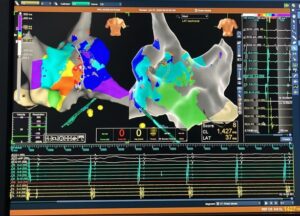
Figure 5. Completed final activation model after ablation, showing an organised slower rhythm on the case display.
What Patients Should Know Before Considering Ablation
Patients often ask whether ablation is safe at an older age. The balanced answer is that age by itself is not the only factor. The decision should be based on symptoms, frailty, heart function, stroke risk, kidney function, lung status, bleeding risk, anaesthesia suitability and personal goals. An 84-year-old who is active and symptomatic may be very different from another patient of the same age with multiple uncontrolled illnesses.
Before ablation, the evaluation usually includes ECG documentation of the rhythm, echocardiography to assess heart structure and function, blood tests, review of medications, stroke-risk assessment, and discussion of anticoagulation. If AFib is intermittent, a Holter monitor, longer patch monitor or smartwatch-supported data may help document the rhythm pattern. Sleep apnoea, obesity, high blood pressure and diabetes should also be addressed because these factors can affect long-term rhythm control.
On the procedure day, patients are generally asked to fast. Sedation or anaesthesia is provided for comfort. Catheters are inserted through the leg vein and guided to the heart. Depending on the case, the team builds 3-D maps, performs pulmonary vein isolation, treats additional circuits if present, confirms endpoints and then monitors recovery. Most patients stay in hospital briefly and return to routine activities within a few days with simple precautions, but the exact timeline depends on the individual case.
Who Should Meet a Heart-Rhythm Specialist?
A consultation with a cardiac electrophysiologist is worth considering if you have repeated palpitations, a racing or irregular pulse, unexplained breathlessness, tiredness that limits routine activity, dizziness, fainting, smartwatch rhythm alerts, AFib that is not controlled on current medicines, or concerns about stroke prevention. Early assessment gives patients a wider range of options, including lifestyle correction, medicines, cardioversion, ablation and long-term follow-up planning.
For patients already diagnosed with AFib, it is also important to ask whether the rhythm is paroxysmal, persistent or long-standing persistent; whether the heart’s pumping function is normal; whether blood thinners are indicated; and whether symptoms are due to AFib or another cause. These answers help personalise treatment.
| When to seek urgent care
Seek urgent medical help for severe chest pain, fainting, stroke-like symptoms, severe breathlessness, or a very fast heartbeat with weakness or low blood pressure. This blog is not for emergency decision-making. Frequently Asked QuestionsIs PFA better than every other ablation method?Not necessarily. PFA is a major advance for selected AFib ablation targets, especially pulmonary vein isolation. But some rhythm problems still need radiofrequency ablation or another strategy. The best method depends on the rhythm diagnosis and map findings. Can AFib come back after ablation?Yes, AFib can recur in some patients. Ablation can reduce arrhythmia burden and symptoms, but long-term success also depends on blood pressure, weight, sleep apnoea, diabetes, alcohol intake, heart structure and follow-up care. Is an elderly patient automatically unsuitable for ablation?No. Age alone is not the only deciding factor. The decision depends on overall health, symptom burden, rhythm type, procedural risk and patient goals. Why was RFA used if the case was a PFA case?PFA was used to isolate the pulmonary veins for AFib. RFA was used because the patient also had a mapped left atrial flutter circuit, which required point-by-point treatment of a critical isthmus. Will I need blood thinners after ablation?This depends on your stroke-risk profile and your doctor’s assessment. Do not stop anticoagulation unless your treating doctor advises it. Suggested Blog Closing / CTAAFib is common, but it should not be ignored. With expert rhythm evaluation, advanced 3-D mapping and the right ablation strategy, many patients can move from uncertainty to clarity. If you or a family member has been diagnosed with atrial fibrillation, or if you experience palpitations, breathlessness, fatigue or an irregular pulse, consider meeting a heart-rhythm specialist for a personalised assessment. For appointments: Dr. M. S. Chandramouli, MD, DM, IDCC – Consultant Cardiac Electrophysiologist, Department of Cardiology, Yashoda Hospitals, Secunderabad. Phone: 7989595637. Appointments: +91 93912 00549. Website: www.rhythmcare.in Short Social Caption for Blog PromotionAtrial fibrillation is more than an irregular heartbeat – it can affect confidence, stamina and quality of life. This case explains how advanced 3-D mapping, pulsed field ablation and targeted radiofrequency ablation were used in a single sitting for persistent AF with left atrial flutter. Read the full patient-friendly explanation by Dr. M. S. Chandramouli, Consultant Cardiac Electrophysiologist, Yashoda Hospitals, Secunderabad. #AtrialFibrillation #AFib #PulsedFieldAblation #PFA #CardiacElectrophysiology #HeartRhythm #YashodaHospitals #DrChandramouli #HyderabadCardiology #HeartHealth Internal Source Notes
Medical DisclaimerThis blog is for general education and awareness only. It is not a substitute for personal medical advice, diagnosis or treatment. Treatment decisions for atrial fibrillation, atrial flutter, anticoagulation and ablation should be made after consultation with a qualified doctor who has reviewed the patient’s individual condition. |