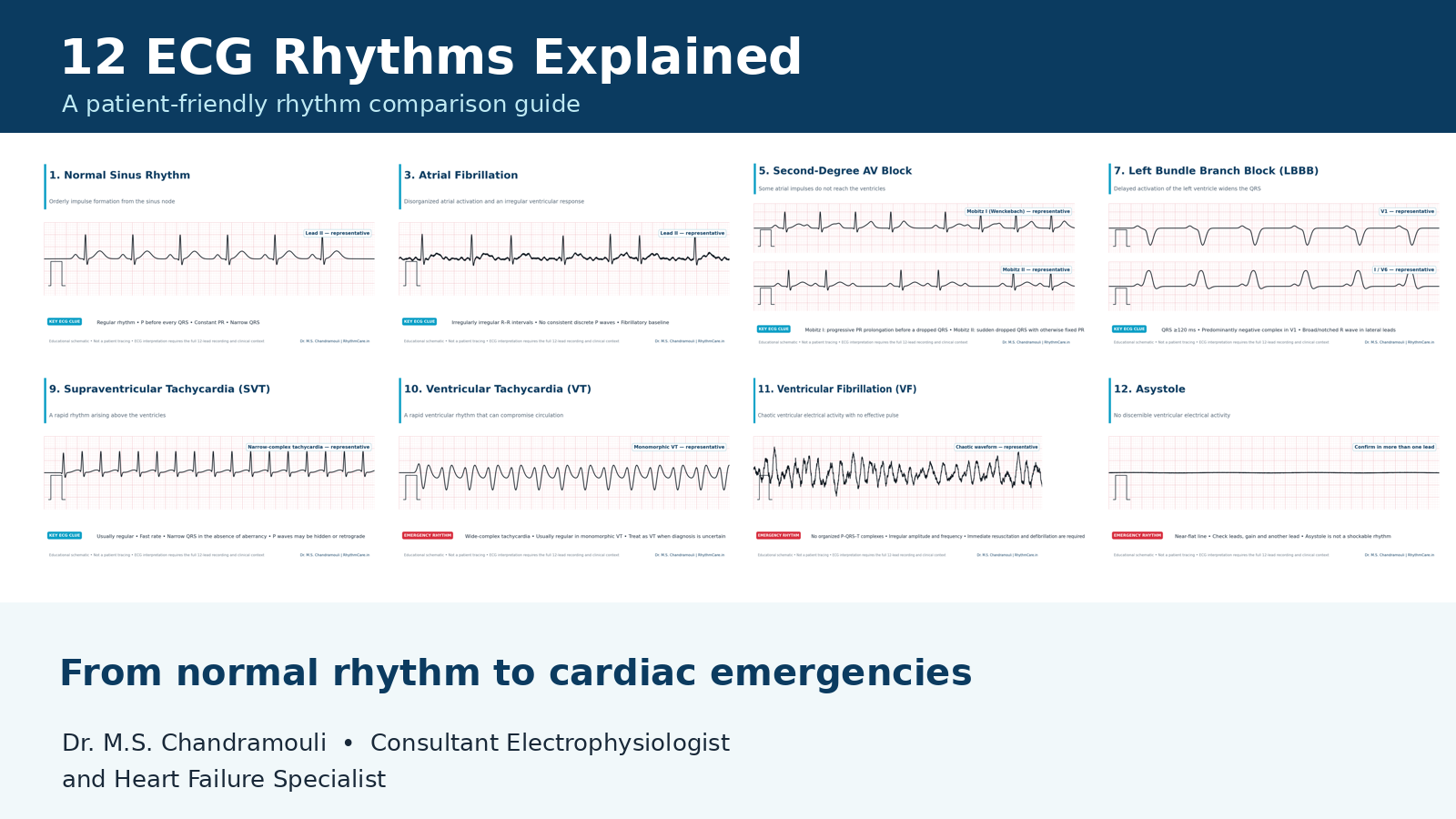
Consultant Electrophysiologist and Heart Failure Specialist, Yashoda Hospitals, SecunderabadAn electrocardiogram (ECG or EKG) records the heart’s electrical activity. These illustrations are educational schematics, not patient tracings, and are not intended for self-diagnosis.
An ECG can reveal whether an impulse begins in the expected place, travels normally through the conduction system and activates the ventricles in an organized way. The tracing must always be interpreted alongside symptoms, medicines, clinical examination and the complete 12-lead recording.
12 ECG patterns at a glance
- Normal Sinus Rhythm
- Atrial Flutter
- Atrial Fibrillation (AF)
- First-Degree AV Block
- Second-Degree AV Block: Mobitz I and Mobitz II
- Third-Degree (Complete) AV Block
- Left Bundle Branch Block (LBBB)
- Right Bundle Branch Block (RBBB)
- Supraventricular Tachycardia (SVT)
- Ventricular Tachycardia (VT)
- Ventricular Fibrillation (VF)
- Asystole
1. Normal Sinus Rhythm
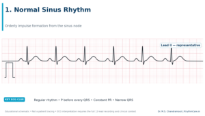
Educational schematic. Not a patient tracing.
What it is
Normal sinus rhythm is the heart’s usual organized rhythm. The electrical impulse begins in the sinus node, travels through the atria, pauses briefly at the AV node and then activates the ventricles through the His–Purkinje system.
What the ECG may show
The rhythm is regular, the adult resting rate is generally 60–100 beats per minute, each QRS is preceded by a similar P wave, the PR interval is constant (usually 120–200 ms), and the QRS is normally under 120 ms.
Why it matters
A tracing that meets these features is reassuring about rhythm origin and conduction at that moment, but a ‘normal ECG’ does not rule out every heart problem. Intermittent rhythm disturbances may require Holter, patch or event monitoring.
What happens next
If palpitations, blackouts, unexplained breathlessness or chest discomfort continue despite a normal office ECG, medical evaluation is still appropriate.
2. Atrial Flutter
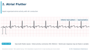
Educational schematic. Not a patient tracing.
What it is
Atrial flutter is an organized but very rapid electrical circuit in the atria, often revolving around the right atrium. The atria may activate around 250–350 times per minute, while the AV node allows only some impulses to pass to the ventricles.
What the ECG may show
Instead of normal P waves, the baseline may show regular ‘saw-tooth’ flutter waves, especially in the inferior leads. The ventricular rhythm may be regular with fixed conduction such as 2:1, or irregular when conduction varies.
Why it matters
Symptoms may include a racing or pounding heartbeat, fatigue, dizziness, breathlessness or reduced exercise tolerance. Like atrial fibrillation, flutter can be associated with clot and stroke risk, so anticoagulation decisions require an individualized risk assessment.
What happens next
Management may include rate control, rhythm restoration and catheter ablation. Typical flutter is often highly amenable to ablation, but the treatment plan depends on the patient’s rhythm mechanism and health.
3. Atrial Fibrillation (AF)
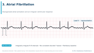
Educational schematic. Not a patient tracing.
What it is
Atrial fibrillation is a common arrhythmia in which atrial electrical activity becomes disorganized. The atria quiver rather than contracting in a coordinated way, and impulses reach the ventricles unpredictably.
What the ECG may show
The hallmark is an ‘irregularly irregular’ ventricular rhythm with no consistent, discrete P waves. Fine or coarse fibrillatory activity may be present, although the baseline can sometimes appear nearly flat between QRS complexes.
Why it matters
AF may cause palpitations, tiredness, breathlessness, dizziness or no symptoms at all. It matters even when silent because it can increase the risk of stroke and can contribute to heart failure or tachycardia-mediated weakening of the heart.
What happens next
Treatment is organized around stroke prevention, symptom control and management of associated conditions. Options can include anticoagulation, rate-control medication, antiarrhythmic medication, cardioversion and catheter ablation after specialist evaluation.
4. First-Degree AV Block
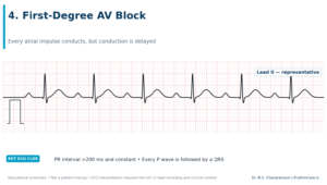
Educational schematic. Not a patient tracing.
What it is
In first-degree AV block, every impulse from the atria reaches the ventricles, but the journey through the AV conduction system takes longer than usual. It is therefore a conduction delay rather than a true dropped-beat block.
What the ECG may show
Every P wave is followed by a QRS, but the PR interval is longer than 200 ms and remains constant from beat to beat.
Why it matters
It is frequently found incidentally and may be related to high vagal tone, medications or conduction-system disease. The significance depends on symptoms, the degree of PR prolongation, other ECG abnormalities and the clinical setting.
What happens next
Many people only need observation and review of contributing medication or conditions. Symptoms such as fainting or marked exercise intolerance call for a more complete rhythm and conduction assessment.
5. Second-Degree AV Block: Mobitz I and Mobitz II
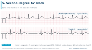
Educational schematic. Not a patient tracing.
What it is
In second-degree AV block, some atrial impulses conduct to the ventricles and others do not. Identifying the subtype is important because the location and risk of the block can differ.
What the ECG may show
Mobitz I (Wenckebach) typically shows progressive PR prolongation until a P wave is not followed by a QRS, after which the cycle resets. Mobitz II shows intermittent non-conducted P waves without progressive PR lengthening; the PR intervals of conducted beats remain similar.
Why it matters
Mobitz I can be benign in selected settings, whereas Mobitz II more often suggests disease below the AV node and may progress to complete heart block. A 2:1 pattern cannot always be safely assigned to either subtype from a short strip alone.
What happens next
New second-degree block, symptoms, a slow pulse or associated bundle branch block requires prompt medical assessment. Pacemaker need depends on the subtype, symptoms, cause and level of block.
6. Third-Degree (Complete) AV Block
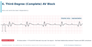
Educational schematic. Not a patient tracing.
What it is
Complete heart block occurs when no atrial impulses conduct to the ventricles. The atria continue under one pacemaker and the ventricles rely on a slower escape rhythm, so the two chambers beat independently.
What the ECG may show
P waves and QRS complexes have no fixed relationship (AV dissociation). The P–P intervals and R–R intervals may each be regular, but the atrial rate is faster. QRS width depends on where the escape rhythm begins.
Why it matters
The slow ventricular rate can reduce blood flow to the brain and other organs, causing fatigue, dizziness, breathlessness, fainting, low blood pressure or cardiac arrest.
What happens next
This can be an emergency. Symptomatic or unstable patients require immediate hospital care and temporary stabilization; a permanent pacemaker is commonly required when the block is persistent and not due to a reversible cause.
7. Left Bundle Branch Block (LBBB)
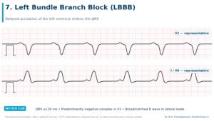
Educational schematic. Not a patient tracing.
What it is
LBBB means conduction through the left bundle is delayed or interrupted, so the left ventricle activates later through cell-to-cell spread from the right ventricle. This changes the sequence of ventricular depolarization and repolarization.
What the ECG may show
Complete LBBB generally has a QRS duration of at least 120 ms, a predominantly negative complex in V1 and broad or notched R waves in lateral leads such as I, aVL, V5 and V6. Secondary ST–T changes are common.
Why it matters
LBBB may reflect underlying structural heart disease, and it can make assessment of ischemic changes more complex. A new LBBB with concerning symptoms deserves urgent evaluation rather than interpretation from the ECG pattern alone.
What happens next
Evaluation often includes clinical examination and echocardiography. Some patients with heart failure, reduced ejection fraction and electrical dyssynchrony may benefit from cardiac resynchronization therapy after guideline-based assessment.
8. Right Bundle Branch Block (RBBB)
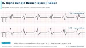
Educational schematic. Not a patient tracing.
What it is
RBBB occurs when electrical activation reaches the right ventricle later than the left. It may be an isolated incidental finding or accompany structural, pulmonary or conduction-system disease.
What the ECG may show
Complete RBBB usually has a QRS duration of at least 120 ms, an rsR′ or terminal R′ pattern in V1 and a broad terminal S wave in leads I or V6. Incomplete RBBB has a similar shape with a shorter QRS.
Why it matters
An isolated RBBB in an otherwise well person may not require treatment. Its importance changes when it is new, occurs with fainting, appears with additional conduction disease or is associated with acute cardiopulmonary symptoms.
What happens next
The ECG should be correlated with symptoms, previous tracings and structural assessment. The bundle branch pattern itself is not treated unless it is part of a clinically significant conduction disorder.
9. Supraventricular Tachycardia (SVT)
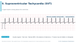
Educational schematic. Not a patient tracing.
What it is
SVT is a group of fast rhythms that arise above the ventricles. Common mechanisms include AV nodal re-entry, an accessory-pathway circuit and atrial tachycardia.
What the ECG may show
Many re-entrant SVTs produce a sudden, regular, narrow-complex tachycardia. P waves may be hidden within the QRS or appear immediately after it. A wide QRS does not exclude SVT, so a wide-complex tachycardia requires expert assessment.
Why it matters
Episodes may start and stop abruptly and cause rapid palpitations, chest awareness, breathlessness, light-headedness or occasionally fainting. Capturing the rhythm during symptoms is extremely helpful.
What happens next
Acute and long-term treatment depends on stability and mechanism. Catheter ablation can provide definitive treatment for many recurrent re-entrant SVTs after electrophysiology evaluation.
10. Ventricular Tachycardia (VT)
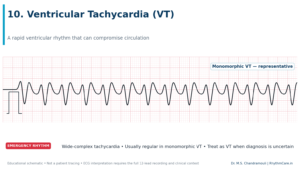
Educational schematic. Not a patient tracing.
What it is
VT is a rapid rhythm originating in the ventricles. It may occur in a structurally normal heart, but sustained VT is often associated with scar, cardiomyopathy, previous heart attack or other structural disease.
What the ECG may show
VT commonly appears as a regular wide-complex tachycardia. AV dissociation, capture beats or fusion beats can support the diagnosis, but absence of these findings does not exclude it. A wide-complex tachycardia should be treated as VT when the diagnosis is uncertain.
Why it matters
Some patients remain awake; others develop chest pain, breathlessness, low blood pressure, fainting or cardiac arrest. VT can deteriorate into ventricular fibrillation.
What happens next
Sustained VT requires urgent assessment. Unstable VT requires immediate synchronized cardioversion; longer-term care may include treatment of reversible triggers, medication, catheter ablation and/or an implantable cardioverter-defibrillator depending on the cause and risk.
11. Ventricular Fibrillation (VF)
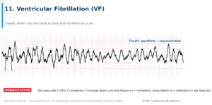
Educational schematic. Not a patient tracing.
What it is
VF is chaotic ventricular electrical activity. The ventricles cannot pump blood effectively, so the person has no effective pulse and rapidly loses consciousness.
What the ECG may show
The tracing shows a continuously irregular waveform with changing amplitude and frequency and no organized P waves, QRS complexes or T waves. Very fine VF can resemble asystole, making lead and gain checks important during resuscitation.
Why it matters
VF is cardiac arrest and is fatal without immediate action.
What happens next
Call emergency services, begin high-quality CPR and use an automated external defibrillator as soon as available. VF is a shockable rhythm; every minute without CPR and defibrillation reduces the chance of survival.
12. Asystole
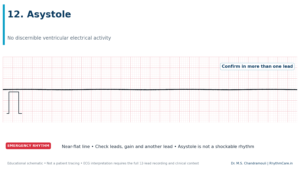
Educational schematic. Not a patient tracing.
What it is
Asystole is the absence of discernible ventricular electrical activity. It represents cardiac arrest, not simply a very slow heart rate.
What the ECG may show
The monitor shows a near-flat line without QRS complexes. Clinicians confirm the rhythm in more than one lead and check electrodes, cables and gain to exclude technical error or fine VF.
Why it matters
There is no effective cardiac output and no pulse. The rhythm requires immediate resuscitation and a search for reversible causes.
What happens next
Call emergency services and start CPR. Asystole is not treated with defibrillation; resuscitation follows cardiac-arrest protocols with high-quality CPR, appropriate medication and correction of reversible causes.
Frequently Asked Questions
Can a smartwatch diagnose an arrhythmia?
Wearables can capture useful single-lead recordings, but they do not replace a clinical 12-lead ECG or specialist interpretation.
Can the office ECG be normal despite palpitations?
Yes. A longer Holter, patch or event monitor may be needed to capture an intermittent episode.
Is a bundle branch block the same as a blocked artery?
No. Bundle branch block concerns electrical conduction; a coronary blockage concerns blood supply to the heart muscle.
About Dr. M.S. Chandramouli
Dr. M.S. Chandramouli is a Consultant Electrophysiologist and Heart Failure Specialist at Yashoda Hospitals, Secunderabad. His focused services include arrhythmia management, RFA and 3D mapping, pacemaker implantation, AICD, CRT and leadless pacemaker procedures.
Consultation
Yashoda Hospitals – Secunderabad
Alexander Road, Kummari Guda, Shivaji Nagar, Secunderabad, Telangana 500003
Phone: 906 342 0686
Website: rhythmcare.in
Medical Disclaimer
This article is for general education and does not replace medical diagnosis or treatment. ECG interpretation requires the full tracing and clinical context.