A Comprehensive Guide to Hardware and Tools for Safe Transvenous Lead Extraction
Cardiology | Electrophysiology | Device Management
Introduction
Cardiac implantable electronic devices (CIEDs)—including pacemakers, implantable cardioverter-defibrillators (ICDs), and cardiac resynchronization therapy (CRT) devices—have revolutionised the management of cardiac arrhythmias and heart failure worldwide. As global implantation rates continue to climb, so too does the incidence of one of the most feared complications: device-related infection. CIED infections represent a life-threatening condition that demands prompt recognition, accurate diagnosis, and definitive treatment to reduce morbidity and mortality.
The cornerstone of managing CIED infection is complete device and lead removal. Transvenous lead extraction (TLE) has emerged as the gold standard approach, supported by international guidelines from the Heart Rhythm Society (HRS), the European Heart Rhythm Association (EHRA), and multiple professional bodies. However, the procedure itself is technically demanding, requiring specialised hardware, experienced operators, and a multidisciplinary team approach. Despite clear guideline recommendations, real-world adherence remains suboptimal—recent studies show that only 24–38% of patients with confirmed CIED infections undergo lead extraction, leaving a significant treatment gap that translates directly into increased mortality.
This blog provides an in-depth exploration of CIED infections, the rationale for lead extraction, and—most importantly—the specialised hardware and tools that make safe, successful transvenous lead extraction possible. Whether you are a clinician, trainee, or healthcare professional seeking to understand this critical field, this guide covers every essential aspect.
Understanding CIED Infections
What Are CIED Infections?
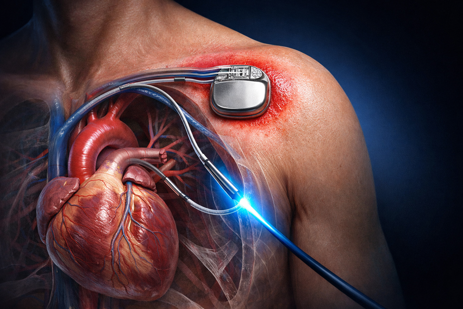
A CIED infection occurs when bacteria colonise the implanted device, its leads, or the surgical pocket in which the generator sits. Once established, the infection can spread along the intravascular leads—effectively using them as a highway into the heart—potentially causing endocarditis, sepsis, and death if left untreated. The risk of infection rises with the duration of device implantation, the number of prior procedures, and patient-specific factors such as immunosuppression, renal failure, and diabetes.
Types of CIED Infections
CIED infections broadly fall into two categories. Pocket infections are localised to the generator site and may present with erythema, warmth, swelling, wound dehiscence, or frank purulence at the pocket. Systemic infections involve bloodstream dissemination and can lead to lead-related endocarditis, with vegetations forming on the intracardiac portions of the leads. Systemic infections carry significantly higher mortality and are frequently caused by Staphylococcus aureus, the most common and aggressive pathogen in CIED infections.
The Clinical Urgency
The data supporting prompt intervention are compelling. Research demonstrates that infection relapse occurs in 50–100% of cases treated with partial device removal or antibiotics alone, compared with relapse rates of only 0–4.2% following complete system removal. Furthermore, lead extraction performed within six days of diagnosis is associated with a 42.9% lower risk of death compared with delayed removal. A seven-fold increase in 30-day mortality has been observed when antibiotics are used with device removal but without lead extraction. These statistics underscore a critical message: delays in referral and extraction cost lives.
Why Lead Extraction Is Essential
International guidelines are unequivocal: complete device and lead removal is a Class I recommendation for confirmed CIED infections. The rationale is straightforward—retained hardware serves as a persistent nidus for infection, making eradication with antibiotics alone virtually impossible once biofilm has formed on the lead surfaces.
A landmark 2025 Australian cohort study of over 2,300 patients confirmed that lead extraction was associated with a 36% reduction in one-year mortality (adjusted hazard ratio 0.64). Despite these benefits, only 24% of infected patients underwent extraction within 30 days. Similar patterns emerge globally: a 2025 Japanese registry analysis found that only 38% of hospitalised patients with CIED infections underwent TLE across all hospitals, though the rate in certified extraction centres rose significantly from 54% in 2015 to 71% in 2021. Independent predictors of not receiving extraction include advanced age, female sex, dementia, cerebrovascular disease, and chronic kidney disease—highlighting the need for improved education and referral pathways.
The Stepwise Approach to Transvenous Lead Extraction
Modern TLE follows a systematic, stepwise escalation protocol. The operator begins with the least invasive technique and progressively advances to more specialised tools as clinical necessity dictates. This graduated approach maximises safety while achieving the highest possible success rate.
Step 1: Direct Manual Traction
The simplest approach involves applying steady traction directly to the lead. For leads implanted less than two years, simple traction alone may be sufficient, as significant fibrotic encapsulation has not yet developed. For active fixation leads, the helical screw is first retracted using a retraction wrench or curved haemostats before traction is attempted.
Step 2: Locking Stylets and Enhanced Traction
When manual traction fails, a locking stylet is inserted into the lead lumen. This device engages the internal coil structure, distributing traction force along the entire lead body rather than concentrating it at the proximal end. This prevents lead fracture and enables more controlled extraction. If traction with a locking stylet remains unsuccessful, the operator escalates to powered dissection tools.
Step 3: Powered Sheaths
Powered sheaths—either laser or mechanical—are advanced over the lead to dissect through fibrotic adhesions binding the lead to the vessel wall and cardiac structures. These represent the workhorses of complex lead extraction.
Step 4: Femoral or Jugular Bail-out
If the superior approach fails, operators may employ a femoral or jugular snare technique to capture and extract the lead from an alternative vascular access point. This multi-access approach can achieve complete success rates exceeding 96%.
Hardware for Lead Extraction: A Detailed Guide
The success and safety of TLE depend heavily on the hardware available to the operator. Modern extraction suites contain a diverse armamentarium of tools, each designed for specific clinical scenarios. Below is a comprehensive breakdown of every major tool category.
1. Locking Stylets
Locking stylets are the foundational tool in any lead extraction procedure. They are thin, flexible wires inserted into the central lumen of the lead being extracted. Once advanced to the lead tip, the stylet is activated to lock against the inner coil, creating a rigid, unified structure that transfers traction force along the full length of the lead. This prevents the lead from stretching, uncoiling, or fracturing during extraction.
Key products in this category include the Liberator Universal Locking Stylet manufactured by Cook Medical, which features simplified sizing and a robust locking mechanism suitable for a wide range of lead types. The Lead Locking Device (LLD) by Spectranetics/Philips offers an alternative approach, locking along the entire lead lumen rather than just at the tip, which provides enhanced force distribution and is particularly useful in older or more fragile leads. Traditional locking stylets required precise sizing to match the lead’s internal diameter; newer designs have simplified this process significantly.
2. Non-Powered (Telescoping) Sheaths
Telescoping sheaths are mechanical, unpowered tools used in a coaxial arrangement to dissect leads free from surrounding fibrotic tissue. They are available in sizes ranging from 7 French to 16 French and are constructed from various materials, each with distinct properties. Teflon sheaths are soft and flexible, ideal for navigating venous curves but less effective against dense scar tissue. Polypropylene sheaths offer greater stiffness and superior scar-disrupting capability, though they require careful handling to avoid vascular injury. Stainless steel sheaths are reserved for the most challenging cases involving dense, calcified fibrosis at the central venous entry points.
The operator advances the inner sheath over the lead body, applying counterpressure to mechanically break apart the encapsulating fibrosis. A larger outer sheath provides additional support and can further dissect in a piston-like action. Non-powered sheaths remain indispensable even in the era of powered tools, as they often complement laser or mechanical sheaths during complex procedures.
3. Excimer Laser Sheaths
Excimer laser sheaths represent one of the most important advances in lead extraction technology. These devices use ultraviolet laser energy (typically at 308 nm wavelength) to dissolve fibrotic tissue through photochemical ablation and photothermal mechanisms. Unlike mechanical dissection, which tears through scar tissue, the laser vaporises adhesions with minimal force, making it particularly valuable for chronic leads with dense, calcified encapsulation.
The GlideLight Laser Sheath by Spectranetics/Philips is the most widely used system, powered by the CVX-300 Excimer Laser generator. The laser fibres are arranged at the tip of the sheath and fire in a circumferential pattern as the sheath is advanced over the lead. The operator simultaneously applies gentle rotation and countertraction to optimise tissue ablation and maintain lead alignment. Sheath sizes are selected based on lead diameter, and fluoroscopic guidance is used throughout to monitor advancement.
While laser sheaths have demonstrated high technical success rates, comparative studies have shown higher major complication rates (approximately 8.4%) compared with mechanical alternatives. This has prompted many centres to adopt mechanical sheaths as first-line powered tools, reserving laser as a crossover option for cases where mechanical dissection alone proves insufficient.
4. Powered Mechanical (Rotating) Sheaths
Powered mechanical sheaths use a rotating threaded or bladed tip to cut through fibrotic tissue as the sheath is advanced over the lead. These tools have become the preferred first-line powered extraction device in many high-volume centres due to their favourable safety profile and comparable efficacy to laser sheaths.
The Evolution and Evolution RL sheaths by Cook Medical feature a bidirectional rotating tip that prevents lead entanglement—an improvement over earlier unidirectional designs. The Evolution RL, introduced in 2019, refined the tip design for less aggressive tissue interaction while maintaining cutting efficiency. The TightRail Rotating Dilator Sheath by Spectranetics/Philips, introduced in 2015, employs a similar rotating mechanism and has shown excellent results in clinical studies. Both systems achieve procedural success rates of 82–86%, comparable to laser sheaths, but with significantly fewer major complications (approximately 1.2% versus 8.4%).
In head-to-head comparisons, when crossover between tools was required, mechanical sheaths used as a second-line tool after failed laser extraction demonstrated superior clinical success compared with the reverse scenario, suggesting that having both tool types available offers the best outcomes in complex cases.
5. Outer Support Sheaths
Outer support sheaths work in tandem with powered extraction tools to enhance their dissecting capability and provide structural stability during challenging extractions. The VisiSheath by Philips and the SteadySheath by Cook Medical are the two primary outer sheaths used in contemporary practice. These larger-bore sheaths are advanced over the inner powered sheath, providing a stable platform that prevents buckling and assists in managing tissue that “snowploughs” ahead of the inner tool. In a piston-like action, the outer sheath can itself contribute to tissue dissection, particularly when encountering highly calcified adhesions.
6. Femoral Workstation and Snare Devices
When the superior approach encounters insurmountable resistance, the femoral approach provides a critical bail-out option. The Byrd Femoral Workstation Sheath by Cook Medical is a curved, deflectable sheath designed to be advanced from the femoral vein into the right atrium and superior vena cava. Used in conjunction with the Needle’s Eye Snare (also by Cook Medical), it allows the operator to capture the distal end of a lead or lead fragment and extract it inferiorly through the femoral access site. This approach is particularly effective for pacing leads and coronary sinus leads, and serves as an essential rescue tool when the primary extraction fails.
7. Bulldog Lead Extender
The Bulldog Lead Extender by Cook Medical is a simple but critical rescue tool. It consists of a metal wire with an end loop through which a disrupted lead can be threaded. When a lead fractures during extraction and the proximal segment separates, the Bulldog can be deployed to grasp the remaining externalised portion, effectively extending the lead to allow continued extraction. An extraction sheath can then be advanced over the lead-Bulldog assembly to complete the procedure.
8. Ancillary and Emergency Equipment
Beyond the extraction-specific tools, a comprehensive lead extraction programme requires extensive ancillary and emergency equipment. Fluoroscopy and transoesophageal echocardiography (TEE) provide real-time imaging during the procedure. A pericardiocentesis tray must be immediately available to manage cardiac tamponade—the most feared complication of TLE. Temporary pacing equipment is essential since extraction removes the patient’s existing pacing system. Cardiopulmonary bypass equipment must be on standby, and a cardiothoracic surgeon must be on-site throughout the procedure. Many centres have developed dedicated “extraction carts” that contain all necessary emergency equipment alongside the extraction tools, ensuring rapid access should complications arise.
Hardware Comparison at a Glance
| Tool Category | Key Products | Mechanism | Best Use Case |
| Locking Stylets | Liberator (Cook), LLD (Philips) | Locks inside lead lumen for full-length traction | All extractions; foundational tool |
| Telescoping Sheaths | Teflon, Polypropylene, Steel (various) | Mechanical counterpressure dissection | Moderate fibrosis; complement to powered tools |
| Laser Sheaths | GlideLight (Philips) + CVX-300 | UV laser photochemical ablation at 308nm | Dense calcified adhesions; crossover tool |
| Mechanical Sheaths | Evolution RL (Cook), TightRail (Philips) | Bidirectional rotating tip dissection | First-line powered tool; chronic leads |
| Outer Sheaths | VisiSheath (Philips), SteadySheath (Cook) | Structural support + piston dissection | Support for powered sheaths in complex cases |
| Femoral Tools | Byrd Workstation + Needle’s Eye Snare (Cook) | Inferior snare capture and extraction | Bail-out; atrial and CS leads |
| Lead Extender | Bulldog (Cook Medical) | Wire loop grasps disrupted leads | Lead fracture rescue |
Outcomes, Safety, and the Future
Contemporary TLE achieves procedural success rates exceeding 94% with major complication rates of approximately 1.7% and procedure-related mortality of 0.3%. The stepwise approach—starting with simple traction and escalating through locking stylets, powered sheaths, and femoral bail-out—maximises both safety and success. A recent analysis of step-by-step efficacy demonstrated that the first-line powered tool achieved success in approximately 65% of cases, rising to 75% after crossover to an alternative tool, 85% with a femoral snare bail-out, and 92% following non-emergency surgery.
Looking ahead, the field continues to evolve with improvements in sheath design, better imaging integration, EMR-based alert systems for early infection identification, and growing recognition of the need for multidisciplinary management teams. The adoption of leadless pacing technologies may eventually reduce the burden of lead-related infections, though the current installed base of traditional CIEDs ensures that expertise in lead extraction will remain essential for decades to come.
Frequently Asked Questions (FAQs)
- What is a CIED infection and how does it differ from a typical surgical site infection?
A CIED infection specifically involves colonisation of the implanted device, its leads, or the generator pocket by microorganisms. Unlike a simple surgical wound infection, CIED infections involve hardware that extends into the bloodstream and heart chambers. Bacteria form biofilms on the lead surfaces that are resistant to antibiotics, which is why complete hardware removal is typically necessary for cure. The infection can manifest as a localised pocket infection or progress to systemic bloodstream infection and even endocarditis with vegetations on the leads inside the heart.
- Why can’t CIED infections be treated with antibiotics alone?
Biofilm formation on the lead surfaces is the primary reason. Once bacteria adhere to the hardware and produce biofilm, they become protected from both the host immune system and most antibiotics. Clinical data show that relapse occurs in 50–100% of cases treated with antibiotics without complete device removal, compared with less than 5% relapse when the entire system is extracted. This is why international guidelines give a Class I recommendation for complete device and lead removal in confirmed CIED infections.
- What is the difference between a locking stylet and a regular stylet?
A regular stylet is a simple wire inserted into the lead to provide stiffness during implantation. A locking stylet, by contrast, is specifically engineered for lead extraction. It has an activation mechanism that causes it to expand, grip, or lock against the inner coil of the lead, creating a rigid connection that distributes extraction force along the entire lead body. This dramatically reduces the risk of lead fracture and allows for safer, more controlled traction during the extraction procedure.
- How does a laser sheath work, and when is it used?
A laser sheath contains optical fibres at its tip that deliver ultraviolet excimer laser energy at 308 nanometres wavelength. This energy dissolves fibrotic tissue through photochemical ablation, vaporising the adhesions that bind the lead to the vessel wall. The laser is activated in short pulses as the operator advances the sheath over the lead under fluoroscopic guidance. It is primarily used for leads with dense, calcified fibrotic encapsulation that cannot be cleared by mechanical dissection alone, and increasingly serves as a crossover tool when first-line mechanical sheaths are insufficient.
- Are mechanical rotating sheaths safer than laser sheaths?
Comparative studies suggest that mechanical rotating sheaths have a more favourable safety profile. One large single-centre study reported major complication rates of 1.2% with rotating mechanical sheaths versus 8.4% with laser sheaths, with comparable procedural success rates around 83–86%. A meta-analysis of over 13,000 patients also found that laser sheath use was associated with increased risk of major complications or death. However, both tools have specific strengths, and having access to both allows for optimal outcomes in the most complex cases through crossover strategies.
- What happens if a lead breaks during extraction?
Lead fracture is a recognised complication, particularly with older or recalled leads. If the proximal lead separates from the intracardiac portion, rescue tools such as the Bulldog Lead Extender can grasp the remaining externalised segment. Femoral snare devices like the Needle’s Eye Snare can be deployed to capture free-floating lead fragments from below. These bail-out techniques are a routine part of extraction planning, and experienced operators are prepared for this scenario with pre-positioned emergency equipment.
- Who should perform transvenous lead extraction?
TLE should be performed by operators with specialised training in lead extraction at centres equipped with the full range of extraction tools and immediate access to cardiothoracic surgical backup. Guidelines mandate that a surgeon proficient in managing potential complications must be on-site during every extraction, and cardiopulmonary bypass equipment must be readily available. High-volume extraction centres consistently demonstrate better outcomes, reinforcing the importance of referring patients to experienced operators.
- What is the role of a multidisciplinary team in managing CIED infections?
Optimal management of CIED infections requires collaboration between electrophysiologists, cardiothoracic surgeons, infectious disease specialists, cardiac imaging experts, and critical care physicians. A multidisciplinary team ensures accurate diagnosis through appropriate imaging and cultures, timely decisions about extraction versus conservative management, selection of appropriate antibiotic regimens, and coordinated planning for device reimplantation when necessary. Institutions that have implemented formal multidisciplinary device infection teams report improved guideline adherence and patient outcomes.
- How long after extraction can a new device be reimplanted?
The timing of reimplantation depends on the type and severity of infection, the causative organism, and the patient’s clinical response to treatment. For pocket infections without bloodstream involvement, reimplantation may occur as early as 72 hours after extraction with negative blood cultures. For systemic infections and endocarditis, guidelines typically recommend completing a full course of intravenous antibiotics (often 4–6 weeks) and confirming infection clearance before reimplantation. The new device should ideally be placed on the contralateral side to avoid reinfection at the original site.
- What advancements are on the horizon for lead extraction technology?
The field is advancing on multiple fronts. EMR-based alert algorithms are being developed to identify infected patients earlier and facilitate timely referrals. Next-generation mechanical sheaths continue to refine tip designs for improved safety and efficacy. Intracardiac echocardiography integration during extraction is improving real-time procedural guidance. Leadless pacing technologies such as the Micra transcatheter pacemaker may reduce future lead-related infection burden, though they do not address the existing population of patients with traditional transvenous systems. Additionally, antimicrobial device coatings and absorbable antibiotic envelopes are being studied for infection prevention at the time of implantation.
Disclaimer: This blog is intended for educational and informational purposes only. It does not constitute medical advice. Healthcare professionals should refer to current clinical guidelines and institutional protocols for patient management decisions.